


Let us help you find what you’re looking for.
Popular Searches
What is - Ventricular Septal Defect
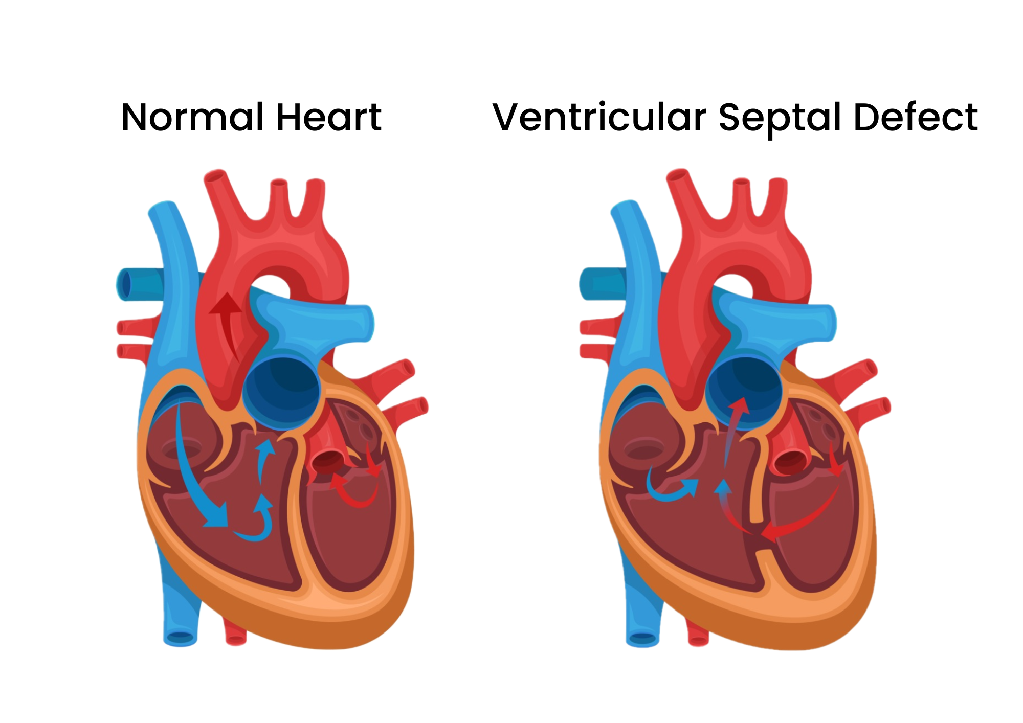
The left and right sided blood circulation are usually kept separate. In patients with VSD, blood from the left lower chamber flows through the hole into the right lower chamber due to a pressure difference.
Ventricular septal defect (VSD) is a congenital defect of the ventricular septum, the wall which separates the heart’s left and right lower chambers (ventricles). During the formation of the heart, at the early parts of pregnancy, the ventricular septum does not fully develop, resulting in a hole. Typically, the left side of the heart pumps oxygen-rich blood from the heart to the rest of the body while the right side of the heart pumps oxygen-poor blood to the lungs. In order to prevent mixing of oxygen-rich and –poor blood, they are completely separated by the ventricular septum. However, when there is a defect/hole between the ventricles, blood from the heart’s left lower chamber is forced through the defect into the right lower chamber because of higher pressure in the left lower chamber. Therefore, a portion of oxygen-rich blood is recirculated back into the lungs resulting in inefficiency and forcing the heart to work harder.
Possible complications of Ventricular Septal Defect (VSD)
Leaving VSD untreated can lead to further complications and the severity of these complications depend on the size of the defect. If the VSD is significant in size, it may lead to the following complications:
- Enlarged heart: Due to the additional blood volume recirculation and increased workload of the heart.
- Pulmonary hypertension: High blood pressure may build up in the lung arteries from the increased blood flow to the lung.
- Heart failure: The heart has to work harder, resulting in a large amount of blood being pumped to the lungs. This can eventually lead to heart failure.
- Abnormal heart rhythms/Arrhythmia: In some cases, VSD might lead to irregular heart rhythms.
Complications associated with smaller defects include:
- Endocarditis: This heart infection is a possible complication of VSD though it is rare.
- Aortic regurgitation: Leakage of blood through the aortic valve.
- Double chamber right ventricle: Abnormal muscle growth in the right ventricle resulting in two separate right ventricular pressure compartments.
Ventricular Septal Defect (VSD) classification
There are various types of VSD and they are classified according to their location within the ventricular setum.
1) Outlet VSD
The hole is located in the “outlet” portion of the ventricular septum, below the aortic and pulmonary valves.
2) Inlet VSD
The hole is located just below the tricuspid valve in the right ventricle and mitral valve in the left ventricle. It could also be a part of an atrioventricular septal defect (AVSD).
3) Muscular/Trabecular VSD
The hole is located in the lower part of the ventricular wall and there is often more than one hole. This is seen in 20% of infants with VSD.
4) Perimembranous/Central VSD
This hole is in the upper section of the ventricular wall.
Symptoms of Ventricular Septal Defect
The symptoms of ventricular septal defect (VSD) may or may not present at birth depending on the size of the hole. If the defect is small, the baby might only show symptoms when they are older or not at all. However, a large defect may cause the baby to experience the following symptoms:
- Shortness of breath or quick breathing
- Sweating
- Fatigue while eating or playing
- Poor appetite or weight gain
Some people with VSD may enter adulthood without knowing about their condition. Adults can also develop VSD due to complications from a previous heart attack or heart procedure, though this is rare. As such, you will need a visit to your doctor as early as possible if you develop the following symptoms:
- Breathlessness
- Palpitations (abnormal sensation of the heart beating)
- Lower limb swelling
- Syncope (fainting)
- Giddiness
- Chest pain
Ventricular Septal Defect - How to prevent
Ventricular septal defect (VSD) is congenital and thus, it is difficult to prevent your baby from having this condition. However, a healthy pregnancy is still crucial to minimising the risks of VSD. Here are some steps you can take:
- Avoid harmful substance like alcohol, tobacco and illicit drugs.
- Make sure you keep up to date with your vaccinations to avoid infections that can be harmful to a developing fetus.
- Speak with your doctor early to get prenatal care and supplements even before you get pregnant. Discuss your current health, lifestyle and medications with your doctor. He/she will then provide you with advice for a healthy pregnancy.
- Eat a balanced diet and exercise regularly.
- Manage diabetes well if you have the condition and work closely with your doctor to keep it under control before getting pregnant.
More importantly, if you have a family history of congenital heart defects or other genetic disorders, speak with your doctor before getting pregnant.
Ventricular Septal Defect - Causes and Risk Factors
Ventricular septal defect (VSD) is a congenital heart defect which means that problems occur during the early stages of the heart’s development. Although genetic and environmental factors have been found to contribute to the presence of VSD, no clear cause of VSD has been found. Generally, genetics are recognised as the biggest risk factor for VSD and it can be associated with other genetic problems as well. In rare cases, VSD can also be developed later in life as a result of a heart attack or previous heart procedures.
Diagnosis of Ventricular Septal Defect
Diagnosis of ventricular septal defect (VSD) normally starts with a physical examination by a doctor, assessing your medical history and routine tests. During the physical examination, the doctor might hear a heart murmur when using a stethoscope. He/she might recommend other tests such as:
- Cardiac catheterisation: A thin, flexible tube (catheter) is inserted into a blood vessel and guided into the heart. This allows the doctor to determine if a VSD is present and whether the heart is functioning well.
- Chest X-ray: This allows the doctor to see if the heart is enlarged or if the lung has excess fluid.
- Echocardiogram: This is an ultrasound scan of the heart that can be conducted on a fetus. It shows any issues with the heart structure. It can also determine the position, size and severity of the VSD.
- Electrocardiogram (ECG): This helps detect any heart defects or abnormal heart rhythms (arrhythmia) by measuring the heart’s electrical activity.
- Cardiac MRI: This allows the doctor to measure the size of the heart and how much blood is flowing across the VSD.
Treatment for Ventricular Septal Defect
There are various ways to treat a ventricular septal defect (VSD) depending on the type, size and severity of the defect. Small VSDs that are not causing symptoms can be left to close on their own though frequent check-ups will have to be conducted to ensure that there are no further complications. However, a larger VSD might require further intervention in the form of medications or surgical procedures. Additionally, some babies with VSD may not feed well and will need additional nutrition to help them grow.
Medications
Medications can be prescribed to treat the symptoms of heart failure that arise due to VSD. These medications generally aim to decrease the amount of blood pumped by the heart and the amount of fluid circulating in the body to prevent a build-up of fluid in the lungs. If pulmonary hypertension develops, targeted medication to lower the pulmonary pressures may be considered.
Surgeries or procedures
Closure of small ventricular septal defect (VSD) may not be required if it does not cause enlargement of the heart. However, closure of a large ventricular defect is recommended to prevent serious problems later in life. Other indications for intervention include infective endocarditis, double chamber right ventricle and progressive aortic regurgitation.
1) Open-heart surgery
This usually involves stopping the patient’s heart temporarily and connecting them to a heart-lung machine which performs the heart’s pumping function. The surgeon will then use stitches or patches to close the hole.
This technique does not involve opening the patient’s chest. The doctor will insert a catheter (thin tube) into a blood vessel and guide it into the heart. The doctor then closes the hole with a specialised mesh device.
After device or surgical closure, the patient will still require regular follow-up by his/her cardiologist.
Ventricular Septal Defect - Other Information
Living with Ventricular Septal Defect (VSD)
Activity
Most patients do not need to restrict their activity. Your doctor will determine if you are required to restrict your activity.
Prevention of Infective Endocarditis (IE)
Antibiotic prophylaxis (taking preventive antibiotics) prior to dental procedures is not needed for patients with unrepaired VSD especially if there are no associated heart defects or complications. However, daily dental hygiene and 6 monthly dental reviews are important for prevention of endocarditis.
After closure of VSD, antibiotic prophylaxis is needed for at least 6 - 12 months depending on whether the defect has completely closed.
Family planning and pregnancy for women with Ventricular Septal Defect (VSD)
Please discuss with your cardiologist regarding birth control methods, pregnancy and prior to starting a family.
Cardiac-Obstetric Clinic
National Heart Centre Singapore (NHCS) and the Department of Obstetrics & Gynaecology from Singapore General Hospital run a monthly specialised joint Cardiac-Obstetric Clinic on the last Monday morning of the month to see all pregnant patients with heart disease. Over the last 10 years, we have managed 200 to 300 pregnant patients with varying severity of cardiac problems through their pregnancy with successful outcomes.
NHCS Adult Congenital Heart Disease (ACHD) Programme
This programme was first started in 2003 and currently offers:
- ACHD clinic on every Wednesday and Thursday afternoon, and on the first and third Fridays of the month in which an average of 20-30 patients are seen at each session. Besides offering care and follow up of ACHD patients, NHCS also does screening of suspected Marfan patients; close monitoring of ACHD patients during pregnancy (see Pregnancy and Women with CHD) and a monthly joint Cardiac Obstetric Clinic (every last Monday of the month) with obstetricians from Singapore General Hospital (SGH) ; Monthly Transition Clinic in KK Women's and Children's Hospital (KKH) run jointly with paediatric cardiologists from KKH for patients above the age of 16. The aim is to give paediatric patients a smooth transition from paediatric cardiology to adult cardiology services. An average of 100-150 teenagers with congenital heart disease are transferred from KKH to NHCS every year. There is also a monthly Pulmonary Hypertension Clinic (every second Friday of the month) run jointly with a respiratory physician and a rheumatologist from SGH for general as well as congenital patients with severe pulmonary hypertension.
- Dedicated twice weekly congenital echocardiography sessions.
- Dedicated weekly congenital cardiac catheterisation and intervention (particularly atrial septal defect (ASD), patent foramen ovale (PFO) and patent ductus arteriosus (PDA) device closure).
- Surgical interventions including extra-cardiac Fontan procedures for uni-ventricular hearts, pulmonary homograft replacements for Tetralogy of Fallot patients with severe pulmonary regurgitation, Rastalli conduit for patients with pulmonary atresia.
- Expertise in nuclear imaging, magnetic resonance imaging (MRI) and multi-slice computed tomography (CT) imaging.
- Expertise in pacing, arrhythmia treatment and ablation in congenital patients.
- Expertise in cardiopulmonary and exercise testing.
- Expertise in heart and lung transplantation for selected patients with end-stage congenital heart disease.
- Dedicated ACHD nurse for education, support and care of ACHD patient and their families.
Adult Congenital Heart Disease (ACHD) Clinic
This clinic looks after patients with operated as well as unoperated congenital heart conditions from age 16 onwards. Adults with congenital heart disease need regular monitoring and sometimes further surgical interventions. It also screens suspected Marfan patients and monitors ACHD patients closely during pregnancy.
For more information on NHCS Adult Congenital Heart Disease (ACHD) Programme, visit here.
Contributed by
The information provided is not intended as medical advice. Terms of use. Information provided by SingHealth.

Our Medical Specialists
Get to know our doctors at SingHealth Hospitals in Singapore.
Get to know our doctors at SingHealth Hospitals in Singapore. here.
Our Medical Specialists
1
2
3
4
5
Health Articles


Stay Healthy The Easy Way
Get trusted health advice, offers and more.
Stay Ahead in Healthcare Industry
Subscribe to our exclusive updates for healthcare professionals.


Keep Healthy With
© 2025 SingHealth Group. All Rights Reserved.