SingHealth Institutions will NEVER ask you to transfer money over a call. If in doubt, call the 24/7 ScamShield helpline at 1799, or visit the ScamShield website at www.scamshield.gov.sg.
Ankle Instability – Diagnosis and Management of Acute and Chronic Ankle Sprains
15 Jul 2020 | Medical News
By Dr Kinjal Mehta, Senior Consultant, Department of Orthopaedic Surgery, Changi General Hospital
A 25-year-old gentleman comes to your clinic with right ankle pain and swelling after an injury two days prior. He was playing soccer and twisted his ankle. He experienced pain and swelling after the incident. He is able to weight-bear on the affected leg. On examination, he has swelling and tenderness overthe lateral aspect of his ankle.
This is a common scenario in GP clinics; the patient has an ankle sprain. With the prevalence of both acute ankle sprains and chronic ankle instability, timely diagnosis and referral is important for effective management.
INTRODUCTION
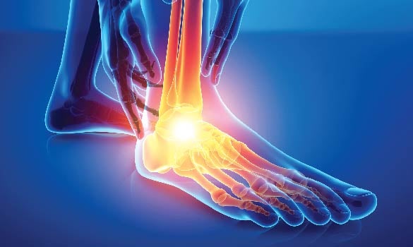
The ankle joint is one of the most commonly injured joints in sports activities and ankle sprain is the most common type of ankle injury1. An incidence rate of 2.15 per 1,000 person years has been reported in the USA2. Ankle sprains commonly occur during sporting activities especially basketball, football and soccer2. They are more common among teenagers, and people in their 20s and 30s.
FUNCTIONAL ANATOMY
The ankle joint consists of the tibial plafond, medial malleolus, lateral malleolus and talus. The main motion of the ankle joint is dorsiflexion and plantar flexion. The talar dome is wider anteriorly than posteriorly. Hence, the ankle joint is more stable in dorsiflexion than plantar flexion. The lateral ankle ligaments are most commonly involved in ankle sprains.
The lateral ankle ligaments consist of the
anterior talofibular ligament (ATFL), calcaneofibular ligament (CFL) and posterior talofibular ligament (PTFL).
The anterior talofibular ligament (ATFL) originates on the anteroinferior border of the fibula and extends to the neck of the talus. It resists anterior translation of the talus in the mortise and is the primary restraint to inversion in plantar flexion.
The calcaneofibular ligament (CFL) originates on the fibula and inserts onto the calcaneus. It is the primary restraint to inversion in neutral or dorsiflexed position.
The posterior talofibular ligament (PTFL) originates on the posterior border of the fibula and inserts on the posterior tubercle of talus. It is the strongest lateral ankle ligament. PTFL limits posterior talar displacement in ankle dorsiflexion.
The most commonly injured ligament is ATFL, followed by CFL and then PTFL. In most patients, PTFL is intact.
CHRONIC ANKLE INSTABILITY
About 30% of patients with acute ankle sprains end up with lateral ankle instability3. Patients with recurrent sprains, a high-grade sprain, ligamentous laxity, lower limb weakness or postural imbalance are more prone to developing chronic ankle instability.
CLINICAL PRESENTATION
1. Acute Ankle Sprains In acute ankle sprains, patients present with pain and swelling over the lateral aspect of ankle after a twisting injury.
On physical examination, tenderness would be palpated on the lateral aspect of the ankle. There should not be any bony tenderness.
The range of motion of the ankle should not be limited. Pain may limit some movement but passive movement should be full.
In the acute setting (one to two weeks) post-injury, physical examination may be difficult due to pain and swelling. The Achilles tendon should be examined to exclude acute Achilles tendon ruptures.
Ottawa ankle rules should be followed to determine if the patient requires x-rays and referral to the emergency department.
2. Chronic Ankle Instability Patients with chronic ankle instability would present with recurrent ankle inversion injuries (sprains).
Patients would usually
describe their symptoms as “ankle giving way” or “ankle feeling loose”. Initially, instability is felt on uneven ground. However, the condition may progress resulting in instability while walking on flat ground.
During an episode of acute sprain, pain and swelling may be present. There should not be any pain or swelling between episodes of sprains.
Physical examination of patients with chronic ankle instability may reveal tenderness on palpation of lateral ankle ligaments.
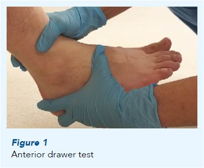
The anterior drawer stress test and talar tilt test may be positive in patients with chronic ankle instability.
The anterior drawer test looks for anterior subluxation of the talus. ATFL can be tested by performing the anterior drawer test in 20 degrees of plantar flexion and comparing it with the uninjured side (Figure 1).
The
talar tilt test is performed by inverting the calcaneus with the foot at 90 degrees. Excessive inversion of calcaneus as compared to the uninjured side indicates injury to CFL
(Figure 2).
Patients with ankle instability may have concomitant osteochondral lesion of talus, peroneal tendon pathology or deltoid ligament injury.
CLASSIFICATION OF ANKLE SPRAINS
Ankle sprains can be graded I-III depending on its severity. Symptoms of swelling, bruising and tenderness increase as the grade of ankle sprain increases. Patients with grade II or III ankle sprains are more likely to have chronic ankle instability.
Grade
Ligament Disruption
Swelling, Ecchymosis,
Tenderness
Pain on Weight-bearing
I
None
Minimal
None
II
Stretch without rupture
Moderate
Mild
III
Complete rupture
Severe
Severe
MANAGEMENT
1. Acute Ankle Sprains Acute ankle sprains are managed using “RICE” (rest, ice, compression and elevation) therapy. Patients are advised to rest and may be given a few days off work depending on their occupation.
Icing the injured ankle will help to decrease symptoms and swelling.
Applying a bandage may also aid in relieving symptoms.
Elevating the injured ankle will help to decrease acute swelling.
The patient may consider wearing a protective brace for comfort. Range of motion exercises should be started from the time of injury.
2. Chronic Ankle Instability 30% of patients with acute ankle sprains may end up with recurrent sprains and chronic lateral ankle instability. Chronic ankle instability is managed with nonoperative measures initially.
Physiotherapy with emphasis on neuromuscular training exercises may result in improvement of symptoms. Peroneal strengthening and balance training using wobble boards are some of the exercises undertaken. Initially isometric exercises are undertaken; these are progressed to dynamic resistive exercises using resistance bands.
Ankle guards or taping especially during sporting activities may help patients manage their symptoms.
Lifestyle changes and activity modifications would also help in controlling symptoms.
Analgesia and non-steroidal anti-inflammatory drugs would help to relieve symptoms after a sprain.
Symptoms may be less pronounced if weight loss is undertaken in overweight individuals.
REFERRAL TO ORTHOPAEDIC DEPARTMENT
Patients should be referred to the emergency department if a fracture is suspected or imaging is required based on the Ottawa rules. Patients with lateral ankle instability should be referred to the orthopaedic department. Initial management would involve nonoperative measures followed by imaging and
surgical management.
IMAGING AND SURGICAL MANAGEMENT
Imaging Stress x-rays using the anterior drawer test or talar tilt test can be performed.
Stress x-rays of the uninjured ankle are also required for comparison.
An ultrasound scan of the ankle ligaments allows real-time assessment of the integrity and laxity of ligaments. A major drawback of ultrasound scans is its operator dependency.
Magnetic resonance imaging (MRI) is usually performed for patients presenting with lateral ankle instability. MRI is useful in evaluating ligament integrity, thickness and bony attachment. MRI can also show concomitant intra- and periarticular pathologies such as osteochondral lesions of talus, tendon injury or ankle impingement.
Surgical Management Surgery is indicated in patients with persistent symptomatic lateral ankle ligament instability after failure of nonoperative treatment.
Surgical management can be divided into anatomic reconstruction and non-anatomic reconstruction of lateral ankle ligaments. Non-anatomic reconstructions (e.g. Chrisman-Snook reconstruction) have fallen out of favour due to complications such as subtalar joint stiffness.
Modified Brostrum-Gould reconstruction is currently considered the “gold standard” operative treatment for lateral ankle instability. Surgery involves anatomica reconstruction of the native ligaments with reinforcement of the inferior extensor retinaculum. Suture anchors may be used to attach ligament to bone.
Anatomical reconstruction with autograft or allograft is usually for patients with insufficient ligament remnants, failed previous lateral ankle ligament reconstruction, high body mass index (BMI) or generalised ligamentous laxity. Ankle arthroscopy may be performed at the time of surgery to address intra-articular pathologies.
Postoperative patients would require six weeks of protected weight-bearing followed by gradual return to activity in a physical therapy programme.
REFERENCES
Fong DT, Hong Y, Chan LK, Yung PS, Chan KM; A systematic review on ankle injury and ankle sprain in sports; Sports Med. 2007;37(1):73-94
Waterman, Brian R.; Owens, Brett D; Davey, Shaunette; Zacchilli, Michael A; Belmont, Philip J. Jr; The Epidemiology of Ankle Sprains in the United States; JBJS: October 6, 2010 - Volume 92 - Issue 13 - p 2279-2284
van Rijn RM, van Os AG, Bernsen RM, Luijsterburg PA, Koes BW, Bierma-Zeinstra SM; What is the clinical course of acute ankle sprains? A systematic literature review; Am J Med. 2008 Apr;121(4):324-331.e6. doi: 10.1016/j.amjmed.2007.11.018.
Dr Kinjal Mehta is a Senior Consultant orthopaedic surgeon and Director of Foot & Ankle Surgery with the Department of Orthopaedic Surgery at Changi General Hospital (CGH). She graduated from the National University of Singapore and did her orthopaedic surgery training in Singapore. Dr Kinjal underwent foot and ankle fellowship training in Canada and has an interest in foot and ankle sports injuries, deformities and trauma. She is also active in teaching medical undergraduates and orthopaedic residents, and attends conferences on a regular basis to present her research.
Medical professionals who would like more information about these conditions, please contact Dr Kinjal Mehta at kinjal.vidyut.mehta@singhealth.com.sg.
GPs can call for appointments through the GP Referral Hotline at 6788 3033.